Core research themes

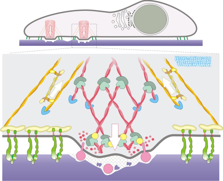
Fig. 1. Representative schemes depicting a cell with podosomes (side view) and below, a single podosome with prominent actin fibers in association with actin regulators (for more details, see Veillat et al., 2015).
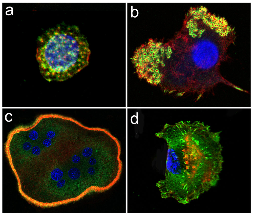
Fig. 2. Immunofluorescent images showing podosomes, seen as red/green dot shaped structures in various kinds of arrangements, in cells from the myelomonocytic lineage (macrophages in a), immature dendritic cells in b) and osteoclasts in c)) and podosome-related structures (invadopodia) in tumor cells (breast cancer cell in d)).
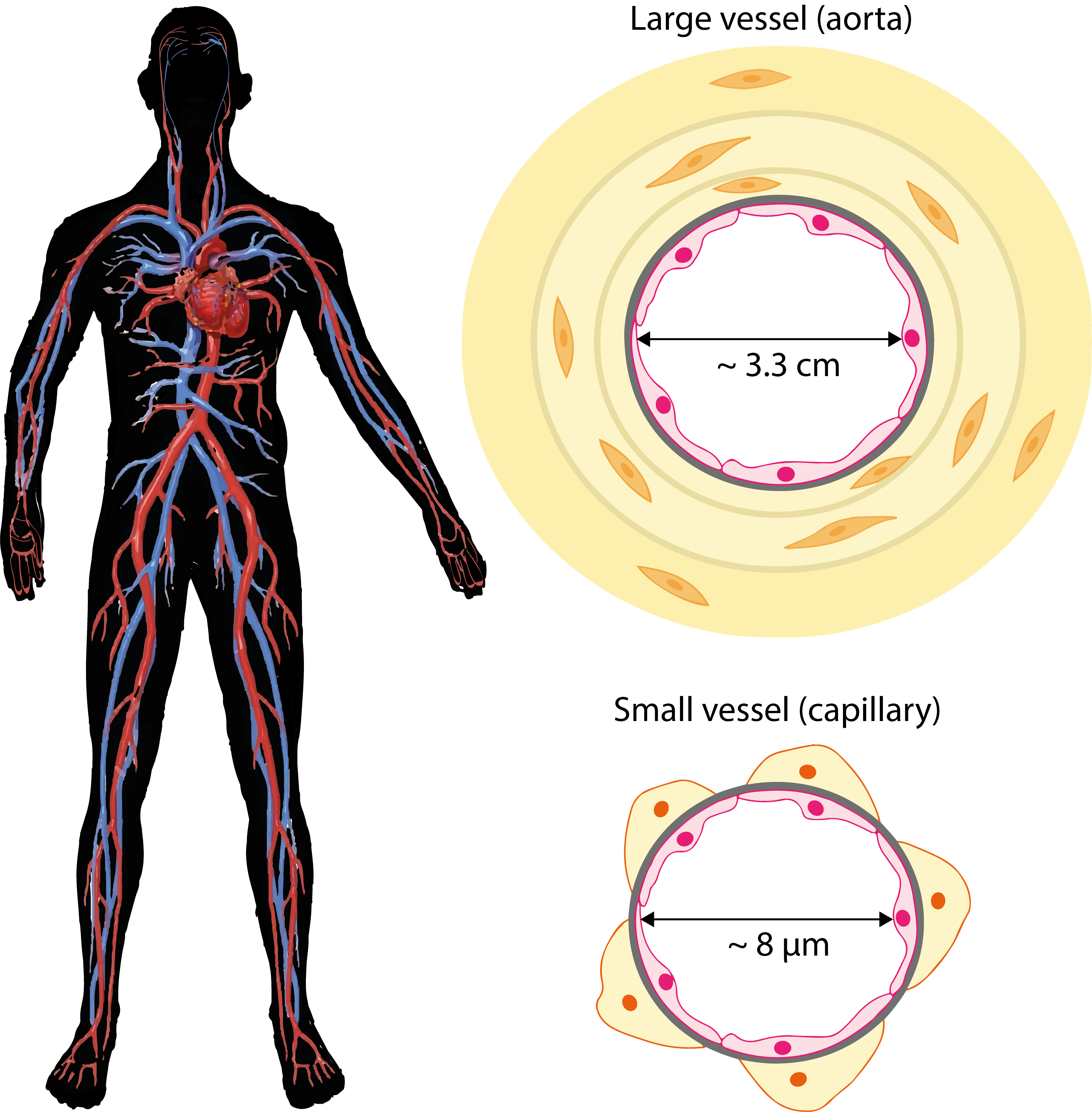
Fig. 3. The vascular system distributes blood throughout the whole body. A diagram of a large artery (like the aorta) and of a capillary (not to the scale), are shown in cross sections on the right.
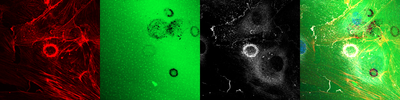
Fig. 4 . An endothelial cell with a podosome rosette degrading the fluorescent gelatin. From left to right are shown: the actin cytoskeleton in red, the fluorescent gelatin in green, a podosome marker (cortactin) in white and the last panel shows the merge of the 3 stainings.
Podosomes are cytoskeletal dot-shaped microdomains (~ 0.5 – 2 mmin diameter) that form at the substrate attached side of certain cells in in vitro cultures. As part of the cytoskeleton, these structures are highly dynamic, they assemble and dissasemble over a minute time scale. By combining adhesion, matrix degradation and mechanosensing capabilities, they enable cells to migrate across tissues.
Structurally, podosomes comprise a concentrated core of actin associated with actin regulatory proteins such as Arp2/3, WASp/N-WASp, cortactin, myosin, dynamin, gelsolin, and cofilin that contribute in their dynamic behavior, and the scaffolding protein and Src substrate Tks5 that constitutes a reliable podosome marker. Around the central core, integrins, adaptors and signaling proteins amongst which FAK, vinculin, talin and paxillin form an adhesive ring domain. Recent studies have now shown that acto-myosin filaments radiating from the dense F-actin cores interconnect podosomes and enable collective behavior. Podosomes also contain metalloproteases (MMPs), transmembrane MMP such as MT1-MMP (MMP14) or membrane associated MMP2 and MMP9, and disintegrin metalloproteinase (ADAMs). With respect to the MMPs. Their presence points towards matrix degradation as the most prominent function of the structures. Thus, MT1-MMP is frequently included as a podosome marker and the ability to proteolyse the underlying extracellular matrix is a key criterion for the identification of podosomes (Fig. 1).
Podosomes are commonly found in cells of the myelomonocytic lineage where they form most of the cytoskeleton (Fig. 2 a-c). They are also found in tumor cells which hijack the podosome-formation machinery to elaborate structures with slightly distinct features, named invadopodia (Fig. 2d). Invadopodia alter cell behavior and enable tumor cells to cross anatomical boundaries.
The vascular system distributes blood throughout the body. Endothelial cells forms a thin monolayer at the luminal side of all blood vessels from the heart to the smallest capillaries.
All vessels, from big arteries to small capillaries, are lined by endothelial cells (that form the endothelium). These cells rest on a basement membrane, a thin layer of extracellular matrix composed of proteins and glycoaminoglycans (shown as a grey circle in Fig. 3). Major protein components are collagen-IV and laminin, that play a role both in attaching the endothelial cells as well as forming a mechanoresistant structure. The basemement membrane is a barrier that contains and separates the endothelium from the rest of the vessel. At the interface of blood and tissues, endothelial cells occupy a strategic position: they perceive and respond to changes in environmental cues such as matrix composition and/or stiffness, blood flow or oxygen concentrations, and they are in direct contact with hormones, growth factors and cytokines that are distributed via the blood.
However, endothelial cells from large and small vessels display distinct morphologies and properties. They respond to distinct signals, consistent with the fact that both types of vessels have distinct structures and functions (Fig. 3). The aorta is the main artery (the largest vessel, 3.5 cm in diameter that carries blood away from the heart to the rest of the body. Capillaries are the smallest blood vessels, their major role is to exchange gases and metabolites of all sorts. Both large and small vessels display a remarkable plasticity; arteries may widen and tiny vessels may lengthen. In all instances, vessel remodeling requires an important change in cell and matrix composition. We have shown that podosomes take central stage in allowing the endothelial cells to breach the basement membrane thus paving the way for remodeling the vascular bed.
We have discovered podosomes in endothelial cells. When plated on a fluorescent gelatin matrix; podosome activity is visualized as black holes corresponding to matrix proteolysis (Fig. 4) and pictured in (Fig. 1). We are exploring their involvement in vascular plasticity.
Experimentally, podosomes are detected by plating the cells on a fluorescent gelatin matrix; podosome activity is visualized as black holes corresponding to matrix proteolysis as shown in Fig. 4 (and depicted in the scheme (Fig. 1)).
Podosomes are found in endothelial cells originating from small and large vessels.